Cardiac Imaging » Echocardiogram

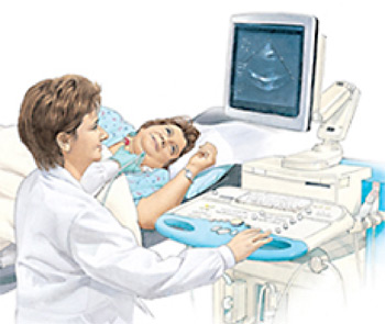
During an echo, images of your heart appear on a monitor.
An echocardiogram (echo) is an imaging test. It helps your doctor evaluate your heart. This test:
- Is safe and painless.
- Can be done in a hospital, test center, or doctor’s office.
- Bounces harmless sound waves (ultrasound) off the heart. A transducer (device that looks like a microphone) is used.
- Helps show the size of your heart. It also helps show the health of the heart’s chambers and valves.
Before Your Echo
- Discuss any questions or concerns you have with your doctor.
- Mention any over-the-counter or prescription medications, herbs, or supplements you’re taking.
- Allow extra time for checking in.
- Wear a two-piece outfit for the test. You may be asked to remove clothing and jewelry from the waist up. If so, you’ll be given a short hospital gown.
During Your Echo
- Most echo tests take 10–20 minutes.
- Small pads (electrodes) are placed on your chest to monitor your heartbeat.
- A transducer coated with cool gel is moved firmly over your chest. This device creates the sound waves that make images of your heart.
- At times, you may be asked to exhale and hold your breath for a few seconds. Air in your lungs can affect the images.
- The transducer may also be used to do a Doppler study. This test measures the direction and speed of blood flowing through the heart. During the test, you may hear a “whooshing” sound. This is the sound of blood flowing through the heart.
- The images of your heart are stored on a computer or recorded on video. This is so your doctor can review them later.
After Your Echo
- Return to normal activity unless your healthcare provider tells you otherwise.
- Be sure to keep follow-up appointments.
Your Test Results
Your doctor will discuss your test results with you during a future office visit. The test results help the doctor plan your treatment and any other tests that are needed.


